The changes that occur to brain structure with normal ageing are likely to impact on cognitive function and neural activity. In order to use this data effectively, the Cam-CAN team are addressing a number of methodological challenges presented by analysis of brain structure across the lifespan.
Current investigations
Currently a number of methodological developments are underway for the Cam-CAN project:
1. Data processing in fMRI: Evaluating segmentation and normalisation methods

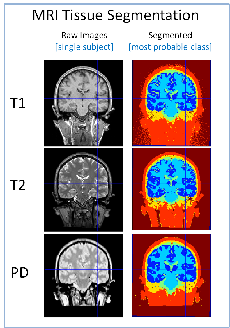
Different MRI sequences have different sensitivity profiles across tissue types, and this has consequences for automated methods that normalise brain images to a standard space and segment them into component tissues (grey matter, white matter, CSF, bone, skin, air). Accurate segmentation/normalisation is
critical for determining the regional integrity of grey and white matter and how these measures change with age (e.g., using VBM); for the statistical analysis of functional MRI images over subjects and across age groups; and for the computation of realistic head models for the estimation of sources of MEG
signals.
In T1-weighted images, signal is high from white matter, intermediate from grey matter, and low from CSF, resulting in the familiar white/grey/black pattern when shown in greyscale (top-left image in figure). In T2-weighted images (middle-left), this pattern is reversed, with high signal coming from CSF,
intermediate signal from grey matter, and low signal coming from white matter. The relative brightness of CSF in T2 images has clinical utility, facilitating the detection of abnormalities such as tumours and white-matter lesions, and may also help separate dura from grey matter during segmentation. Proton-density weighted (PD) images (bottom-left) are useful for extracting the inner surface of the skull and outer surface of the skin for use in MEG
head modelling.
We are evaluating methods for segmentation and normalisation using these different MR contrast images for use in VBM, fMRI, and MEG analyses. Preliminary results suggest that the simultaneous segmentation of all three images (using SPM’s multi-channel segmentation plus normalisation) may provide the most consistent results (e.g., across sessions).
2. DTI and DKI

The Cam-CAN project will use a number of methods to examine white matter integrity, primarily DTI and DKI. Diffusion Tensor Imaging (DTI) is a diffusion MRI modality that became an established technique for studying brain microstructure in several areas such as cerebral ischemia, traumatic brain injury, multiple sclerosis and Alzheimer’s disease, as well as brain maturation and ageing. Diffusion Kurtosis Imaging (DKI) is an extension of DTI which has been shown to provide more robust diffusion measures and values of kurtosis are believed to provide an index of tissue barrier microstructure complexity. From the diffusion and kurtosis tensor, one can compute several invariant parameters such as the mean diffusivity MD and kurtosis MK, which give the average of
diffusion and kurtosis over all 3D spatial directions within a voxel, or indices of tissue anisotropy as the fractionally anisotropy FA. The latter is close to one for tissue consisting of oriented microstructures, as the case of white matter, or close to zero for media where microstructures do not seem to be spatially oriented, as the case of gray matter.
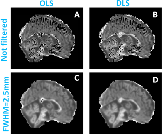
Our aim is to develop robust and accurate procedures for DTI and DKI and use them to analyse brain connectivity changes during ageing. This has involved in part improving the fit achieved with ordinary linear squares (OLS) method (see Figure left, panel A) by using a newly proposed fitting method named direct linear least squares (DLS), which improves the fit (see Figure below, panel B), and by introducing a Gaussian kernel in our pre-processing steps,
the non-biological values along the cortex are removed (panel C). Finally, combining the Gaussian filter with an optimised parameter estimation procedure, results in images no longer show implausible values of kurtosis (panel D).
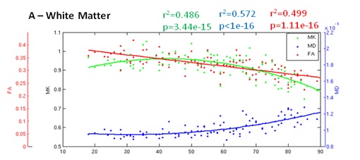
A preliminary analysis of ageing effects examined prefrontal cortex as a region of interest. As reported in previous studies, FA in WM decreases with age which can be a consequence of degeneration of oriented structures i.e. decrease in tissue anisotropy. The processes that influence such decrease may include demyelinisation of axonal fibres and axonal loss. FA is not sensitive to changes in gray matter due to the isotropic nature of this type of tissue. Our MK data suggests a later decrease in MK reflecting a global degeneration of the prefrontal brain.

These preliminary results shown that DKI can provide useful information on structural brain connectivity. However since DKI is a very recent MRI modality, further analysis will be preformed to validate the aging changes measured by the parameters extracted from Kurtosis tensor and to understand better which physiological mechanisms are behind this changes. Our future steps will include the analysis of other brain regions by using different ROIs in addition to the prefrontal ROI or voxel based approaches such as VBM and TBSS, and the study of relationships between the brain microstructure changes and changes in cognitive ability as quantified by the behavioural measures of memory, attention, emotion, language, and action.

 Cambridge Centre for Ageing and Neuroscience